Confocal laser scanning microscopeConfocal laser scanning microscopy
Exterior
no image

no image

Overview
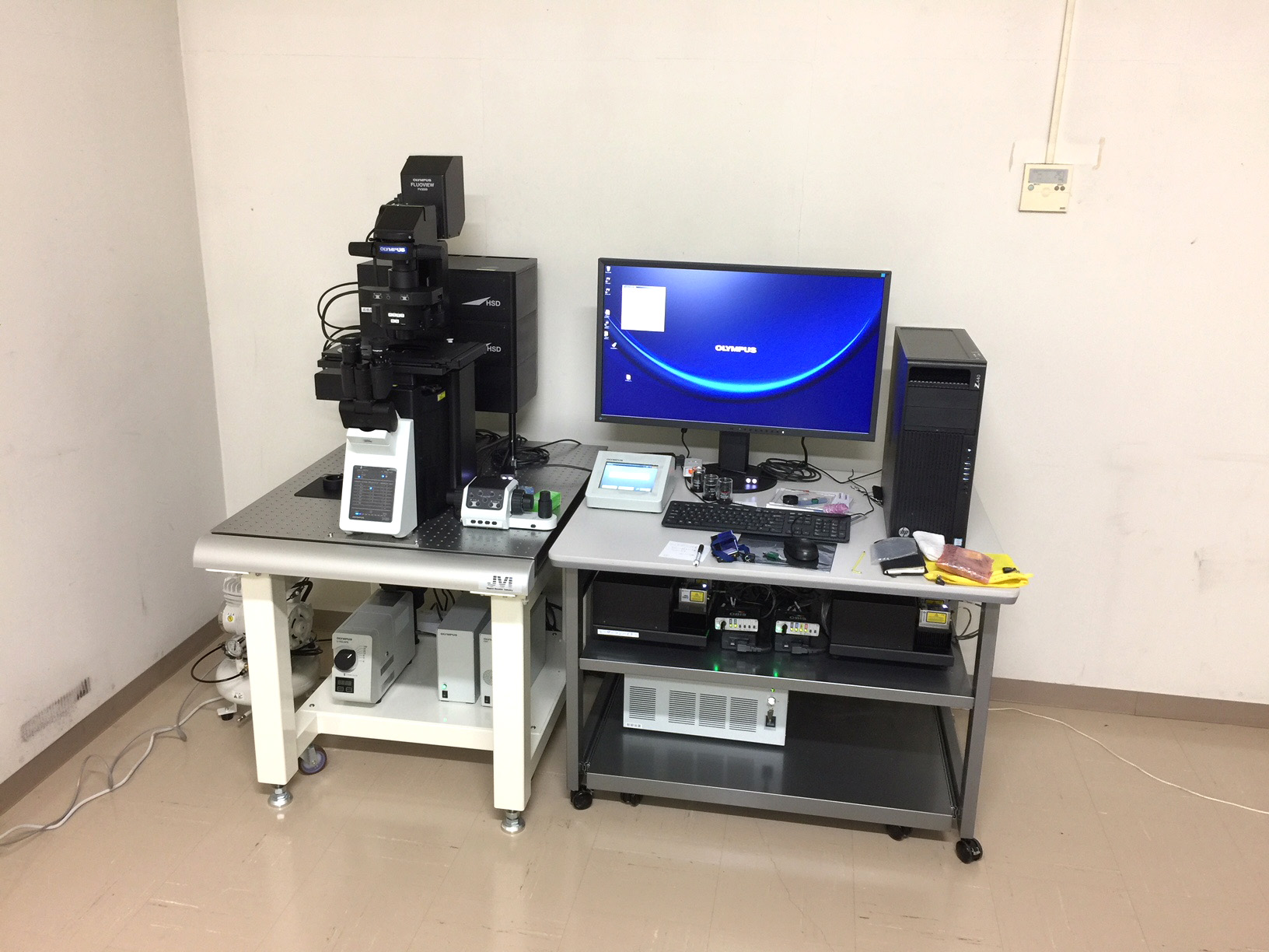
A confocal laser scanning microscope (hereafter abbreviated as CLSM) uses a laser light source to irradiate a sample to be observed with light that has passed through a pinhole. It is a microscope that captures and visualizes with a highly sensitive detector. With ordinary microscopes, light from outside the focal plane impairs the sharpness of the image, but CLSM observes light only from the focal plane, so it is possible to obtain extremely high-resolution and clear images. In addition, by scanning laser light and reconstructing an image with a computer, an optical tomographic image can be obtained, and a three-dimensional image can also be constructed. Such images cannot be obtained with a normal fluorescence microscope. CLSM is used not only for functional analysis at the protein and cell level, but also for a wide range of observations, including tissues and small animals.
In addition, this instrument is a CLSM that has advanced operability so that imaging of living cells and tissues that require high sensitivity and high speed and complicated experimental operation procedures can be easily set. For live imaging and time-lapse observation of living cells, high-sensitivity observation with weak excitation light that does not cause photobleaching and high-speed imaging functions are required. This instrument has sufficient performance to meet these requirements, making it possible to easily observe the elementary processes of cell division and proliferation. In addition, it is possible to obtain high-resolution fluorescence images at the individual and tissue level by precisely synthesizing the field images of the observation target using a computer-controlled motorized stage. Furthermore, the spectral unmixing function enables the overlapping fluorescence spectra to be separated with high precision, enabling the analysis of intracellular protein positional information. This instrument achieves an extremely high resolution of 120 nm using super-resolution technology, and can be used for advanced intracellular molecular imaging.
| Name (abbreviation) | Confocal laser scanning microscope |
|---|---|
| Model number (manufacturer) | FV3000(Olympus) |
| laser combiner | 405nm:50mW 488nm:20mW 561nm:20mW 640nm:40mW |
| Microscope | Inverted Research Microscope Minimum feed step 1㎛ |
| Spectral detector | ・Cooled GaAsP photomultiplier 2CH ・Multi Alkaline Photomultiplier 2CH |
| Scanner | Two silver-coated galvanometer scanner mirrors |
| Installation location | Room ④, 2nd floor, Building C3 |
| Equipment manager | Department of Biochemistry Kenichiro Hayashi |
Contact us
Please feel free to contact us from the following.
