Confocal laser scanning microscopy
OutlineOutline
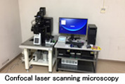
Confocal laser scanning microscopy (CLSM) is one of the most important device in the field of fluorescence imaging and has been considered as an essential tool in biological research. CLSM provide the fluorescent image is obtained from the focal plane only, and CLSM also provide the 2D and 3D digital scanned images constructed by localized laser excitation. CLSM is widely used to study the functional analysis of protein at subcellular level and at tissue and individuals.The FLUOVIEW FV3000 CLSM is suitable for the high sensitivity and speed required for live cell imaging as well as deep tissue observation, the FV3000 CLSM enables a wide range of imaging modalities, including macro-to-micro imaging, super resolution microscopy, and quantitative data analysis. FV3000 is ideal CSLM for developmental biology, stem cell research, electrophysiology, cancer research, slide imaging, and more. The deconvolution algorithm in FV3000 enables overlapping spectra and Fluorescence cross-talk can be eliminated by the unmixing algorithm during live imaging. FV3000 equipped the Olympus Super Resolution imaging module that can acquire fluorescent signals with a resolution of approximately 120 nm and provide the ultra-high resolution image of subcellular compartments.

PerformancePerformance
| Name (abbreviation) | Confocal laser scanning microscopy |
| Model number (manufacturer) | FV3000(Olympus) |
| Laser combiner | 405nm:50mW 488nm:20mW 561nm:20mW 640nm:40mW |
| Scanner | 2 silver-coated galvanometer scanning mirrors |
| Spectral detector | Motorized volume phase holographic transmission diffraction grating, motorized adjustable slit selectable wavelength bandwidth: 1–100 nm, wavelength resolution: 2 nm |
| Microscope | Motorized inverted microscope IX83 (IX83P2ZF) |
| Installation location | Room 4, 2nd floor, Building C3 |
| Equipment manager | Department of Biochemistry Hayashi Kenichiro |
| Contact us | Contact form is open 24 hours a day. Contact by telephone: Direct 086-256-8473 ext.3242 |